What it is:

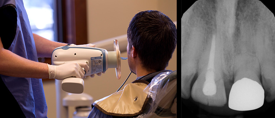
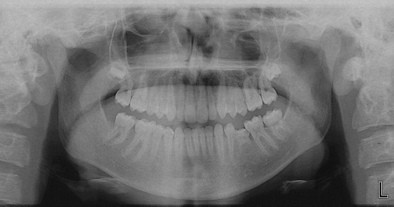
A form of x-ray imaging using sensors instead of traditional photographic film. Digital dental radiographs can be taken inside (intraoral) or outside (extraoral) the mouth. Intraoral x-rays, the most commonly taken dental x-ray, provide great detail and are used to detect cavities, check the status of developing teeth and monitor teeth and bone health. Dental x-rays are one of the lowest radiation dose studies performed. A routine exam which includes 4 bitewings is less than one day of natural background radiation. Digital radiography is an excellent option for those who take x-rays on a regular basis or for those who are concerned about radiation.
An x-ray may reveal:
- small areas of decay between the teeth or below existing fillings
- infections in the bone
- periodontal disease
- abscesses or cysts
- developmental abnormalities
- some types of tumors
Patient Benefits:
Digital x-rays expose patients and staff to up to less radiation than traditional film x-rays, they also have the following additional benefits: They are faster, more comfortable, can be transferred electronically for seamless diagnosis with other care providers and specialists without loss of information and they are better for the environment because they don’t require developing chemicals. They can also be viewed on a computer monitor within seconds of taking the image and diagnosis of problem area is quicker.
Things to Remember:
The frequency of getting x-rays of your teeth often depends on your medical and dental history and current condition. Some people may need x-rays as often as every six months; others with no recent dental or gum disease and who visit Dr. Zaugg regularly may get x-rays only every couple of years.


